baby chest x ray technique
The initial view is from the front and the second is a side view. A chest radiograph for a 12-year-old female is an embarrassing ordeal.

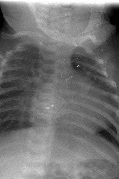
An Ap Erect Chest Xray Of The Patient Showing An Upward Tilted Cardiac Download Scientific Diagram
The umbilical stump remains in situ for approximately 1-2 weeks and.
. Baby chest x ray technique. However all children are modest to some degree about having their genitals or backsides exposed after ages 4 to 5. Correct technique is essential to generating effective percussion notes.
We use the central ray CR tox point the -ray beam where we want it to go. Sometimes other picture views also are taken. The chest radiograph is the most common radiographic procedure performed in the imaging department and is the initial imaging modality in a patient presenting with thoracic symptoms.
When the chest radiograph also includes the abdomen look out for the umbilical clip. AP and lateral oblique views if requested Sacrum and coccyx. Exposure chart was developed with two distinct groups of exposure.
AP lateral and odontoid Thoracic. Pediatric Chest Screen 70-80 DIGITAL OPTIMUM kVp Universal CR Technique Chart using a standard 21 LgM Part View kV mAs kV mAs kV mAs Abdomen AP Grid 85 10 -15 85 20 - 25 85 30 - 40 Ankle AP 70 18 70 2 70 25 Ankle Obl 70 16 70 18 70 22 Ankle Lat 70 15 70 16 70 2 Chest -Adult AP 400 - tt -72 85 2 - 25 85 32 - 4 90 5 - 64. Radiographs of the chest and the abdomen are the most commonly requested diagnostic X-ray examinations undertaken in neonatal intensive care units.
Lateral cervical spines are taken at 150 cm. Chest PAAP erect 180 cm. The degree of rotation is best assessed by comparing the length of the anterior ribs visible on both sides.
Most neonatal chest X-rays are AP films unless the baby is made to lie prone Lucency of soft tissue shadow - darker the soft tissue more is the exposure Ease of visibility of retrocardiac vertebrae if the retrocardiac vertebrae are easily seen the film is over exposed Relative lucency of lung fields. 851a a Use automatic exposure control 500 speed for chestabdomen else 400 speed at specified kVp when practical. Chest x-ray is the most commonly used imaging exam for evaluating the chest.
PA and LATERAL Chest AP neck. As newborn chest radiographs are taken in the AP plane the. These are plastic clips used to clamp the umbilicus before it is cut at birth.
Erect chest X-rays are taken at 180 cm. Chest AP erect in chair 180 cm. X-ray plate - avoid direct contact with baby use X-ray tray provided with the open care system cover with sterile plastic sheetenvelope Avoid direct contact with the cold X-ray plate Provide extra heat source as the open care heater is moved away from the baby or tilt its direction towards baby if possible.
All distal extremity exposures are taken at 110115 cm SID. Take film during inspiration with mouth closed. Frequently for a single child both radiographs are requested simultaneously.
901a a Use automatic exposure control 500 speed for chestabdomen else 400 speed at specified kVp when practical. The Federal Food Drug and. Full legfull spine imaging is performed at 180 cm using CR.
Optimisation strategies body exposures for head trunk humerus femur and. The X-ray film is also developed and viewed with transmitted light on a light box or computer screen. Take film during phonation or cryinginfants.
This is partly due to modesty but also due to fear. Angled AP and lateral. In contrast most 12-year-old males have little modesty about their chests.
Syndromes that may impact the respiratory system. It is often the first type of imaging used to identify sources of pain evaluate traumatic injuries and locate a foreign body. These images can be obtained either as two separate exposures one of the chest and one of the abdomen or as a single.
Make use of digital radiography dr and needle phosphor computerised. Below is a non-exhaustive list of clinical syndromes which can be associated with respiratory system pathology. X-rays are used throughout the body.
This technique represents the expansion in two dimensions only. The abdomen radiograph is a commonly requested examination in the pediatric patient. X-ray examination of KUB on infants - AP Abdominal X-ray.
Normal Anatomy and Artefacts. The chest X-ray technique in young children involves two views. The anteroposterior AP diameter of the neonatal chest is almost as great as its transverse diameter giving the chest a cylindrical configuration.
It can help diagnose and assess. Label films. Children that present for abdominal x-rays are often very unwell therefore specialized techniques and appropriate communication are essential for gaining the childs cooperation.
In pediatric imaging the anteroposterior supine chest x-ray is beneficial for imaging unconscious or uncooperative patients. Medical X-ray imaging has led to improvements in the diagnosis and treatment of numerous medical conditions in pediatric patients. Aerationofthenormalneonatallungisvirtuallycomplete within two or three respiratory cycles after birth and the lung fields should appear symmetrically aerated on the initial X-ray with the diaphragms lying at the level of.
The normal neonatal chest X-ray. Most neonatal chest X-rays are. Chest X-rays can be done while a child is standing sitting or lying down.
X-ray Imaging for Pediatrics. From the back of the chest if the child is old enough to stand up for the X-ray from the side. Usually the X-ray technician will take pictures of the chest.
For younger children the technician will take pictures from the front of the chest and the side. In young children the patient lies on the table and the hands are held above the head. If concerned about consolidation or pneumothorax.
X-ray exams are used to help diagnose a wide variety of injuries and illnesses in children.
Chest X Ray Cxr Congenital Children S Heart Centre

Osteopoikilosis Spotted Bone Disease Radiology Imaging Radiology Bone Diseases

The Double Bubble Sign Is Seen In Infants And Represents Dilatation Of The Proximal Duodenum And Stomach It Radiology Radiology Imaging Radiologic Technology

X Ray Radiologic Technology Child Life Specialist

Chest X Ray Of A 6 Month Old Child With An Icd The Active Can Is Download Scientific Diagram

Pediatric Chest Horizontal Beam Lateral View Radiology Reference Article Radiopaedia Org

A Chest X Ray With The Typical Appearance Of Meconium Aspiration Syndrome Download Scientific Diagram

Human Bones Realistic X Ray Shots Human Bones Human Skeleton For Kids X Ray

Pin On X Ray

Pediatric Chest Supine View Radiology Reference Article Radiopaedia Org

Congestive Heart Disease The Image Shows The Radiograph Of An Infant Who Presented On The Third Day Of Li A Day In Life Congestive Heart Failure Heart Disease

The Normal Cxr Nurse Radiology Nursing Education

Mastoid Ap Axial Towne Methode Radiology Schools Radiology Cassette

Boning Up On Humerus Clavicle And Ac Joint Positioning Radiology Diagnostic Imaging Medical Knowledge

4 Health Benefits And Risks Of X Rays No 1 Unique Dr Heben Human X Ray Walk In Clinic

Pin On Thorax

Getting A Chest X Ray X Ray Radiology Ray

Paratracheal Lymphadenopathy Radiology Case Radiopaedia Org Radiology Radiology Imaging Radiography

Abdominal X Ray Radiological Signs Part I Radiology Abdominal X Ray